
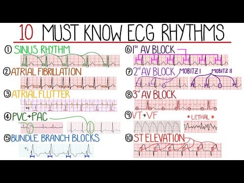
This blog post explores the most common ECG patterns, including sinus rhythm, atrial fibrillation, atrial flutter, premature contractions, bundle branch blocks, AV blocks, ventricular tachycardia, ventricular fibrillation, and ST elevation, providing detailed descriptions and clinical significance of each pattern.
Electrocardiograms (ECGs) are vital tools in medicine for diagnosing various heart conditions. This post will delve into some of the most common and must-know ECG findings, providing a comprehensive overview of each pattern.
Sinus rhythm is the normal rhythm of the heart characterized by regular P waves followed by a QRS complex. This pattern indicates normal conduction of electrical signals from the sinoatrial node in the right atrium through the atrioventricular node and bundle of His into the ventricles. The PR interval, which is the time between the start of the P wave and the initiation of the QRS complex, remains constant and typically ranges from 80 to 100 milliseconds in adults.
Atrial fibrillation is characterized by abnormal electrical activity leading to uncoordinated contractions of the atrial muscle fibers. This results in an ECG with no clear P waves, and QRS complexes are not consistently preceded by P waves. The rhythm is described as irregularly irregular, with variable intervals between the R waves. Atrial fibrillation can be classified based on heart rate:
Often mistaken for atrial fibrillation, atrial flutter features coordinated electrical activity in the atria due to a re-entry circuit. This leads to rapid atrial contractions, typically around 300 beats per minute, and a regular atrial rate. The ECG shows a characteristic sawtooth pattern, particularly in the inferior leads. The ratio of P waves to QRS complexes can indicate the degree of block through the atrioventricular node, with a common ratio being 2:1.
Premature contractions are common findings on ECGs and can be classified into:
Bundle branch blocks can be classified into left and right types, with the left bundle branch further splitting into anterior and posterior branches. These blocks are characterized by wide QRS complexes (greater than 120 milliseconds).
AV blocks are categorized based on the degree of conduction delay:
These two arrhythmias are critical to recognize:
While not an arrhythmia, ST elevation is a significant ECG finding often associated with acute myocardial infarction. It is defined as the portion between the end of the S wave and the beginning of the T wave. The elevation is typically more than one millimeter from the isoelectric baseline in most leads, with specific criteria for leads V2 and V3. Different leads represent various territories of the heart, and ST elevation can indicate the area affected by a blocked coronary artery.
Understanding these common ECG patterns is crucial for healthcare professionals in diagnosing and managing cardiac conditions. Each pattern provides valuable insights into the heart's electrical activity and can guide appropriate treatment strategies. For further reading and resources, consider exploring specialized literature and educational videos on ECG interpretation.