Proteomics is a valuable tool for understanding the fundamental characteristics of an organism through the study of its proteins. This blog post will delve into the intricate processes involved in proteomics, particularly in the context of aquaculture, highlighting the steps from sample collection to protein analysis.
The Importance of Proteomics in Aquaculture
Proteomics allows researchers to gain insights into the biological functions and interactions of proteins within aquatic organisms. By studying proteins, scientists can better understand the health, growth, and overall biology of fish, which is crucial for improving aquaculture practices.
Sample Collection for Proteomics
The first step in proteomics involves the careful collection of samples from fish. Here’s how it is typically done:
- Anesthetizing the Fish: Begin by anesthetizing the fish in a beaker containing phoxyanol at a concentration of 500 ppm. This is suitable for fish averaging 30 grams.
- Measuring and Weighing: Once the fish is anesthetized and turned upside down, remove it from the beaker, weigh it, and measure its dimensions.
- Types of Samples: Various samples can be used for proteomics, including fluid samples like mucus or plasma obtained from blood centrifugation, as well as tissue samples. For tissue samples, a sterilized scalpel is used to open the fish and remove the visceral area, selecting the desired organs for analysis.
- Freezing Samples: Immediately place the selected organs in liquid nitrogen and label them appropriately to preserve their integrity for further analysis.
For this discussion, we will focus on liver tissue extraction, although other tissues can also be utilized. The extraction process involves several key steps:
- Weighing the Tissue: Start by weighing 50 mg of liver tissue from each sample.
- Dissolving the Tissue: Dissolve the tissue in 500 µL of dig buffer, adding 5 µL of protease inhibitor cocktail and 2 µL of EDTA (250 mM).
- Homogenization: Add metal beads to the tubes and homogenize the samples using a tissue laser for two cycles of 30 seconds at a frequency of 25 per second.
- Incubation and Centrifugation: Incubate the homogenized samples at 4°C for 30 minutes with constant rotation. Then, centrifuge at 13,000 g for 15 minutes at 4°C to remove insoluble material.
- Collecting the Supernatant: After centrifugation, collect the resulting supernatant and dilute it 1:180 with the initial buffer.
Protein Quantification
To determine the protein concentration of the diluted samples, a Bradford assay is performed:
- Preparing the Microplate: Add 5 µL of seven curve standards and two controls (blank and dich) in triplicates to a 96-well microplate, along with 5 µL of each sample.
- Adding Reagents: Next, add 250 µL of Bradford reagent to each well and measure the absorbance at 595 nm.
- Estimating Protein Concentration: Use the absorbance readings to estimate the protein concentration and calculate the volume of sample needed to collect 500 µg of protein for subsequent analysis.
To purify the protein extracts from non-protein contaminants, a cleanup kit is utilized:
- Adding Precipitating Agents: Add 300 µL of precipitating agent 1 to the protein sample, vortex, and incubate on ice for 15 minutes.
- Centrifugation: Add 300 µL of precipitating agent 2 and centrifuge at over 12,000 g for 5 minutes to form a tight pellet, discarding the supernatant.
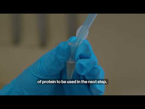
- Washing the Pellet: Add 40 µL of wash reagent 1 on top of the pellet and centrifuge again under the same conditions. Discard the wash and add 25 µL of ultra-pure water, vortexing the mixture.
- Final Steps: Add 1 mL of Bradford wash reagent and 5 µL of wash 2 additive, vortex for 1 minute, incubate at -20°C for 30 minutes (vortexing every 10 minutes), and finally centrifuge for 5 minutes at maximum speed. Remove the supernatant and air dry the pellet at room temperature.
Protein Digestion and Analysis
The resulting pellets are then resuspended, and the proteins are digested into peptides using enzymes such as trypsin. The analysis of these peptides is conducted using advanced mass spectrometry techniques:
- Nanoflow Liquid Chromatography Tandem Mass Spectrometry: This technique separates, ionizes, and analyzes the peptides with high sensitivity.
- Data Matching: The resulting data is matched against protein databases for accurate protein identification, providing insights into the protein composition of the samples.
Conclusion
Proteomics plays a crucial role in advancing our understanding of aquatic organisms in aquaculture. By employing detailed methodologies for protein extraction, quantification, purification, and analysis, researchers can uncover vital information that can lead to improved practices in fish farming and aquaculture management. The integration of omics technologies in this field promises to enhance the sustainability and efficiency of aquaculture operations, ultimately benefiting both the industry and consumers.