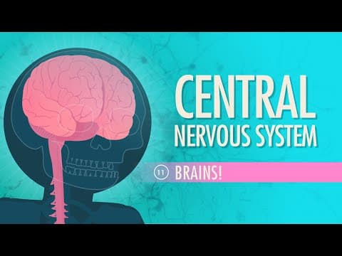
This blog post explores the central nervous system's structure and function through the story of James, a stroke survivor who experienced Broca's aphasia. It details the brain's development, its specialized regions, and how injuries can reveal its complexities, emphasizing the importance of both the central and peripheral nervous systems in human function.
James was a healthy professional, a father of two, and an active member of his community. He enjoyed telling jokes and playing softball on Sundays. However, at the age of 45, his life took a drastic turn when he suffered a stroke. Although he recovered quickly, he faced a significant challenge: he could no longer speak. This condition, known as Broca’s aphasia, resulted from damage to a specific area in the left hemisphere of his brain called Broca’s area, which is crucial for language production and processing.
Broca’s aphasia often leaves individuals with the ability to understand speech but an inability to produce intelligible words. For instance, James could comprehend his wife asking if he wanted cereal for breakfast but could only respond by repeating the word "too." Despite this limitation, he could still intonate as if he were speaking full sentences.
Interestingly, after some time and therapy, James discovered that he could communicate through singing. This unexpected development highlights a fascinating aspect of brain function: singing engages a different region of the brain, located on the right side, which is analogous to Broca’s area on the left. Through practice, James learned to sing words and eventually relearned how to speak by training the right side of his brain to articulate language.
James's story illustrates the brain's vulnerabilities and the specific functions of its various regions. The nervous system is divided into two main networks: the central nervous system (CNS) and the peripheral nervous system (PNS). The CNS consists of the brain and spinal cord, while the PNS comprises the nerves that extend from the CNS throughout the body.
The primary role of the CNS is to integrate sensory information collected by the PNS and respond accordingly, coordinating both conscious and unconscious activities. For example, if the sun is bright, you might shade your eyes; if you're hungry, you might call for pizza. The brain processes all these sensations, thoughts, and directives, while the spinal cord conducts two-way signals between the brain and the rest of the body, managing basic reflexes that do not require conscious thought.
The brain and spinal cord are composed of delicate, jelly-like nervous tissue that is highly susceptible to injury. To protect this vital organ, the brain is encased in the bones of the skull and vertebrae, surrounded by protective membranes known as meninges, and bathed in cerebrospinal fluid. This fluid not only cushions the brain but also allows it to float within the skull, reducing its weight and enabling movement.
Understanding the brain's structure requires a look at its development. In a developing embryo, the CNS begins as a simple neural tube. The lower end of the tube elongates to form the spinal cord, while the upper end expands into three primary brain vesicles: the prosencephalon (forebrain), mesencephalon (midbrain), and rhombencephalon (hindbrain). By the fifth week of development, these vesicles evolve into five secondary vesicles, which lay the groundwork for the adult brain's major regions.
The prosencephalon divides into the telencephalon and diencephalon, while the rhombencephalon forms the metencephalon and myelencephalon. The mesencephalon remains undivided. These vesicles develop into the brainstem, cerebellum, diencephalon, and cerebral hemispheres, each responsible for different functions.
The brainstem, which includes the midbrain, pons, and medulla oblongata, regulates essential involuntary functions such as heart rate, breathing, and sleep. The midbrain is particularly important for processing sensory information and coordinating reflexive motor responses. For example, it allows you to react quickly to a loud sound or track a moving object with your eyes.
The cerebellum, on the other hand, is primarily involved in coordinating muscular activity and balance.
The diencephalon houses structures like the thalamus and hypothalamus, which regulate homeostasis, alertness, and reproductive activities. It also contains part of the limbic system, responsible for strong emotions such as fear. This area is sometimes referred to as the "reptilian brain" due to its shared characteristics with simpler animals, focusing on instinctual behaviors like eating and mating.
The telencephalon undergoes the most significant changes during brain development, forming the cerebrum, which is the largest part of the brain. The cerebrum is divided into two hemispheres, covered by the cerebral cortex (gray matter) and the inner white matter. It governs voluntary movements, complex thinking, learning, emotional regulation, and consciousness.
The cerebral cortex is highly folded, with gyri (ridges) and sulci (grooves) that increase its surface area. Each hemisphere is divided into lobes, each with distinct functions:
James's experience with Broca's aphasia and the subsequent recovery through singing exemplifies the brain's remarkable adaptability and the importance of understanding its structure and functions. The central nervous system, with its intricate organization and specialization, plays a crucial role in our daily lives, from basic reflexes to complex cognitive tasks. As we continue to explore the nervous system, we will uncover more about how it connects with the peripheral nervous system and the broader implications for human health and behavior.