
This blog post explores the inferior view of the skull, detailing important structures such as the foramen magnum, carotid canal, jugular foramen, and various bones and processes. It provides insights into their functions and the anatomical significance of each opening, aiding in the understanding of cranial anatomy.
The inferior view of the skull reveals a complex arrangement of bones and openings that play crucial roles in the anatomy of the human body. This post will delve into the significant structures visible from this perspective, including the foramen magnum, carotid canal, jugular foramen, and various bones and processes.
In the inferior view, one can observe the palatine process of the maxilla and the palatine bone. The palatine process is often confused with the palatine bone itself, as they are closely related yet distinct structures. The palatine process is part of the maxilla, while the palatine bone is a separate entity.
The occipital bone, marked in orange, features horizontal bony projections known as the inferior and superior nuchal lines. These ridges serve as attachment sites for muscles located at the back of the skull. The external occipital protuberance, located centrally, is another important landmark for muscle attachment.
The inferior view of the skull is characterized by several foramina (openings) that allow for the passage of nerves and blood vessels. Here are the major foramina to note:
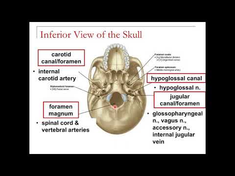
The largest opening in the skull is the foramen magnum, which allows the spinal cord to pass through as it transitions into the brainstem. Additionally, the vertebral arteries also traverse this opening, making it vital for both nervous and vascular systems.
Located on either side of the foramen magnum, the carotid canal (or carotid foramen) is a round opening through which the internal carotid artery passes. This artery is crucial as it supplies blood to the brain, branching from the common carotid artery that runs up the neck.
Just posterior to the carotid canal is the jugular canal (or jugular foramen), which is wider and accommodates multiple structures. The internal jugular vein, which carries deoxygenated blood from the brain back to the heart, passes through this opening. Additionally, three cranial nerves—the glossopharyngeal nerve, vagus nerve, and accessory nerve—also traverse the jugular canal.
The occipital condyles are rounded projections located on either side of the foramen magnum. They articulate with the first cervical vertebra (C1 or atlas), allowing for head movement. Just superior to the occipital condyles is the hypoglossal canal, through which the hypoglossal nerve passes, innervating the muscles of the tongue.
Other openings include the foramen spinosum and foramen ovale, which are smaller and less prominent. The foramen spinosum is associated with the middle meningeal artery, while the foramen ovale allows passage for the mandibular nerve, a branch of the trigeminal nerve.
The vomer bone, which forms part of the nasal septum, is visible in the inferior view. It has a plough-shaped appearance and works in conjunction with the perpendicular plate of the ethmoid bone. The palatine bone, which contributes to the posterior roof of the mouth, is also visible, with the maxilla forming the anterior portion.
The pterygoid processes, which project downward from the sphenoid bone, serve as attachment sites for the pterygoid muscles involved in mastication (chewing).
Understanding the inferior view of the skull is essential for students of anatomy and medicine. By familiarizing oneself with the various openings and structures, including the foramen magnum, carotid canal, jugular canal, and associated bones, one can gain a deeper appreciation for the complexity of cranial anatomy. Sketching these structures and their relationships can further aid in mastering this challenging aspect of human anatomy.